
Martha E. Shenton, PhD

Contact
shenton@bwh.harvard.eduWebsite
http://pnl.bwh.harvard.eduBiography
Dr. Shenton graduated Durant Scholar (Summa cum laude), Phi Beta Kappa, from Wellesley College (1973) where she was a psychology major. She received a Master's degree from Tufts University in 1976 in Developmental Psychology. She then worked as a mental health worker and then as a research assistant, prior to entering the Ph.D. program in Psychology at Harvard University (1978-1984). At Harvard, she worked with Professor Philip Holzman. Upon completing her Ph.D., she entered the Clinical Research Training Program in Biological Psychiatry as a Post-Doctoral Fellow to work with Dr. Robert McCarley. Work with him focused on event related potentials and formal thought disorder but soon involved evaluating the brain using CT and MRI. She began working in the Surgical Planning Laboratory with Drs. Ferenc Jolesz and Ron Kikinis in 1988. Drs. Shenton, McCarley, Kikinis, and Jolesz have been actively collaborating on investigating brain abnormalities in schizophrenia using MRI since 1988. Dr. Shenton remained a member of Dr. McCarley's laboratory from 1984 to 2005, where she was appointed Post-Doctoral Fellow (1984-1986), Instructor (1986-1989), Assistant Professor (1989-1993), Associate Professor (1993-2000), and Professor (2000) in the Department of Psychiatry, first at Massachusetts Mental Health Center (1984-1985) and then at the VA Boston Healthcare System, Harvard Medical School. In 2003, Dr. Shenton was also appointed as Professor of Radiology, Brigham and Women's Hospital, Harvard Medical School.On October 1, 2005, Dr. Shenton accepted a new appointment as Professor in the Department of Psychiatry, Brigham and Women's Hospital, and Director of the newly founded Psychiatry Neuroimaging Laboratory. She has received numerous awards and grants for her work in schizophrenia, including a Career Scientist Development Award (K01, 1988-1993), two Independent Scientist Awards (K02, 1994-1999 and 1999-2004), and, a Senior Scientist Award (K05, 2004-2009) from the National Institute of Mental Health (NIMH). She has also received continuous R01 grant support from NIMH for her imaging studies in schizophrenia (since 1992), as well as support from VA Merit Awards. She is co-PI on a VA Schizophrenia Center grant, and she is PI on an Imaging Core and a Project as part of a Boston CIDAR Center grant that is focused on evaluating vulnerability to progression in schizophrenia (2007-2012). She is also PI of the schizophrenia core of a U54 grant entitled "National Alliance of Medical Imaging and Computing", funded by the National Institute of Health. This grant's aim is to develop neuroimaging tools to understand better brain abnormalities in schizophrenia. Additionally, Dr. Shenton has received numerous private foundation grants including two Milton Foundation Awards, two Scottish Rite Awards, a Stanley Foundation Award for Research into Serious Mental Illnesses, as well as being a Senior Mentor for the Stanley Medical Research Institute to support hands-on research experience for high school students, undergraduate students, graduate students, and medical students in the field of mental health (1997-2004). Since 1999, her research focus has also included diffusion tensor imaging of white matter fiber tracts in schizophrenia. She is author on more than 200 peer reviewed empirical articles and proceedings, as well as author or co-author on multiple book chapters. She is Associate Editor of Brain Imaging and Behavior, and she has been on the editorial board of Schizophrenia Bulletin and is currently on the editorial board of Schizophrenia Research, and Psychiatry Research: Neuroimaging, as well as being a frequent reviewer for another fifteen journals. She has served on the National Institute of Health Study Committees, including the following Initial Review Groups: Clinical Neuroscience and Biological Psychiatry, Brain Disorders and Clinical Neuroscience-6, Conte Center grant reviews, and the Neural Basis of Psychopathology, Addictions and Sleep Disorders Study Section. She has also reviewed grants for the Wellcome Trust in the United Kingdom and for the March of Dimes Birth Defect Foundation. She also serves as a member of the Board of Honors Tutors in the Department of Psychology (Faculty of Arts and Sciences), Harvard University, Cambridge, MA, and she has been Chair and Co-Chair of the Research and Development Committee, VA Boston Healthcare System, as well as for the Department of Psychiatry at Harvard Medical School, and she serves as a member of the Subcommittee of Professors, Harvard Medical School. She has received numerous awards throughout her career, including the fifth recipient of the Joseph Zubin Memorial Fund Award for Research in Psychopathology.
Research Summary
Research work involving the investigation of brain abnormalities in schizophrenia has progressed considerably over the past decade and a half largely due to the improved spatial resolution of MR images and to new image processing tools used to extract information from MR scans. We now know that the temporal lobe, particularly the amygdala-hippocampal complex, parahippocampal gyrus, and superior temporal gyrus, are implicated in the pathophysiology of schizophrenia. These brain regions are also correlated with gray matter volume reductions in prefrontal cortex, suggesting that brain regions involving temporal-frontal connections may be importantly involved in schizophrenia.Our work has now led us to evaluate patients early in the course of their illness, as well as to evaluate individuals with schizotypal disorder who share some of the features common to schizophrenia but without psychosis, and without some of the confounding effects observed in schizophrenia studies such as medication and duration of illness (chronicity). We have also expanded our work to fMRI to evaluate both cognitive and structural brain regions involved in schizophrenia, and we have also taken advantage of a recent technology, diffusion tensor imaging, to evaluate white matter fiber tracts in the brains of psychiatric patients and controls.
Our work has also led us to work on problems involving the coregistration of multimodal imaging techniques such as MRI, fMRI, and transcranial magnetic stimulation (TMS). Such studies should further our understanding of schizophrenia, a disorder that not only afflicts just under 1% of the general population, but which also accounts for the use of more hospital beds than any other health problem other than diseases associated with aging.
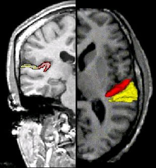
Image is a 1.5 mm slice through the brain. On the Left is a coronal slice where the planum temporale (Yellow) and Heschl's gyrus (Red) are outlined. Planum temporale is an important substrate of Language processing while Heschl's gyrus is involved in auditory processing. The Right side of the image is an axial slice showing the same structures but in 3D.
Selected Publications
- Shenton ME, Kikinis R, Jolesz FA, Pollak SD, LeMay M, Wible CG, Hokama H, Martin J, Metcalf D, Coleman M, McCarley RW. Left temporal abnormalities in schizophrenia and thought disorder: A quantitative MRI study. N Engl J Med 1992;327(9):604-612.213-223.[ESI Thomson Scientific-rated as 4th most cited paper in schizophrenia, from more than 24,000 authors and 19,000 papers, in the decade of the 1990's (http://www.esi-topics.com/schizophrenia/index.html.) (PubMed ID: 1640954)
- Potts GF, Gugino LD, Leventon ME, Grimson WE, Kikinis R, Cote W, Alexander E, Anderson JE, Ettinger GJ, Aglio LS, Shenton ME. Visual hemifield mapping using transcranial magnetic stimulation coregistered with cortical surfaces derived from magnetic resonance images. J Clin Neurophysiology 1998;15(4):344-350. (PubMed ID: 9736468)
- Kwon JS, McCarley RW, Hirayasu Y, Anderson JE, Fischer IA, Kikinis R, Jolesz FA, Shenton ME. Left planum temporale volume reduction in schizophrenia. Arch Gen Psychiatry 1999;56(2):142-148. (PubMed ID: 10025438)
- Niznikiewicz MA, Donnino R, McCarley RW, Nestor PG, Iosifescu DV, O'Donnell BF, Levitt JJ, Shenton ME. Abnormal angular gyrus asymmetry in schizophrenia. Am J Psychiatry 2000;157(3):428-437. (PubMed ID: 10698820)
- Shenton ME, Dickey CC, Frumin M, McCarley RW. A review of MRI findings in schizophrenia. Schizophr Res 2001;49(1-2):1-52.[ESI Thomson Scientific, rated as one of the top 1% of fast breaking papers in the field: http://www.esi-topics.com/fbp/comments/october02-MarthaShenton.html] (PubMed ID: 11343862)
- Gilbertson MW, Shenton ME, Ciszewski A, Kasai K, Lasko NB, Orr SP, Pitman RK. Smaller hippocampal volume predicts pathologic vulnerability to psychological trauma. Nat Neuroscience 2002;5(11):1242-1247. (PubMed ID: 12379862)
- Kubicki M, Westin C-F, Maier SE, Frumin M, Nestor PG, Salisbury DF, Kikinis R, Jolesz FA, McCarley RW, Shenton ME. Uncinate fasciculus findings in schizophrenia: A magnetic resonance diffusion tensor imaging study. Am J Psychiatry 2002;159(5):813-820. (PubMed ID: 11986136)
- Nakamura M, McCarley RW, Kubicki M, Dickey CC, Niznikiewicz MA, Voglmaier MM, Seidman LJ, Maier SE, Westin C-F, Kikinis R, and Shenton ME. Fronto-temporal disconnectivity in schizotypal personality disorder: A diffusion tensor imaging study. Biol Psychiatry 2005;58(6):468-478. (PubMed ID: 15978550)
- Nakamura M, Nestor PG, McCarley RW, Levitt J, Hsu L, Kawashima T, Niznikiewicz M, Shenton ME. Altered orbitofrontal sulco-gyral pattern in schizophrenia. Brain 2007;130:693-707. (PubMed ID: 17347256)
- Bouix S, Martin-Fernandez M, Ungar L, Nakamura M, Koo M-S, McCarley RW, Shenton ME. On evaluating brain tissue classifiers without a ground truth. Neuroimage 2007;36(4):12071224. (PubMed ID: 17532646)