|
|
Welcome to CarDiaNet
The incidence of metabolic disease (obesity, metabolic syndrome, diabetes) is increasing in the developed world, and contributes to the already significant burden of cardiovascular disease. Network researchers point out that not only do metabolic diseases increase the risk of heart disease, but heart disease usually involves some form of change in the baseline metabolism of heart cells, along with an increase in apoptosis (programmed cell death). They hypothesize that networks of signalling exist within the heart, and that problems with these networks can lead to harmful changes in metabolism and inappropriate cell death, which appear clinically as heart failure. One of the major goals of the network is to examine how the pathways for heart cell metabolism and survival are linked, particularly in heart failure. For example, a general resistance in the body to the affects of insulin can diminish the ability of heart cells to repair protect or repair themselves, a condition which would lead to worsening heart failure. At the same time, it has been discovered that heart failure, of itself, can cause cells in the heart to be resistant to insulin, which then worsens the effect. Research into this complex process will yield new insights into cardiac disease related to metabolic disease, and into heart failure generally, and could provide new targets for therapy in a disease that is particularly difficult to treat.
Place your mouse over the images on the left to enlarge.
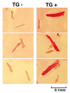
Cardiomyocytes isolated from chimeric hearts of X-linked Akt1 transgenics (right panels) compared to those from littermate controls. Transgene (stained red) expressing cardiomyocytes are larger than non-expressing cardiomyocytes isolated from the same hearts.
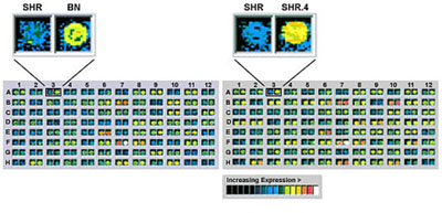
Microarray-based identification of Cd36 as an aberrantly-expressed gene in SHR. The hybridisation signal for SHR is drastically reduced compared to the Brown Norway (BN) and SHR.4 (congenic) control strains. (Aitman et al. (1999). Nature Genetics 21, 76-83.)
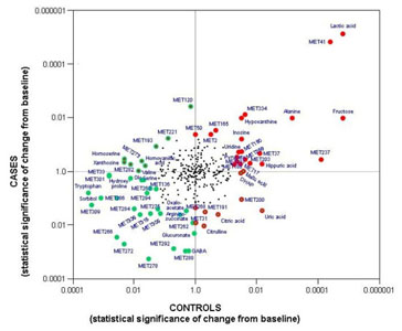 Metabolomic assessment of exercise
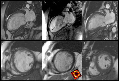
CMR cine stills (panels a and d), and late gadolinium enhancement images (panels b and e) of a lady with severe 3 vessel CAD and heart failure before bypass surgery. The inset (panel e) shows her preoperative PET scan where the numbers express segmental glucose utilization (segments >0.25 mmol/min/g are considered viable). Her post-operative CMR scan at 6 months is shown in panels c and f.
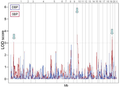
Quantitative trait loci on chromosomes 1, 10, and 19 associated with systolic blood pressures (SBP) measured by radiotelemetry in recombinant inbred strains.
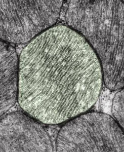
Electron microscopy image of healthy mitochondria in a muscle cell. A single mitochondrion has been digitally colored for clarity. The mitochondria consist of two lipid membranes, the inner and outer membranes. Cell respiration is located in the inner membrane.
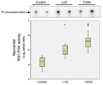
Activation of phosphatidylinositol 3-kinase (PI3K) in heart samples from patients with left ventricular dysfunction (LVD) or type 2 diabetes mellitus (T2DM) as compared to controls.
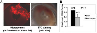
Preliminary findings: Ischemia/reperfusion of the rat heart after delivery of PGC-1a. Rats hearts were injected with adenovirus encoding for PGC-1a. 2 days later, the coronary artery was transiently ligated for 30 minutes, during which time fluorescent microspheres were injected systemically. 24 hours later, hearts were harvested, stained with TTC, and imaged. A: Sample transverse slice of a heart. Left panel shows microsphere fluorescence; non-labeled regions represent areas not perfused during ligation period. Right panel shows TTC staining of the same heart slice; TTC stains live tissue red. B: 2 and 3 rats were injected with adenovirus encoding for GFP and PGC-1a, respectively. The bar graph indicates the fraction of the area at risk (AAR, as determined by microsphere fluorescence) that developed myocardial infarction (as determined by TTC staining).
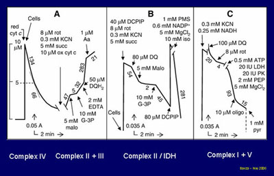
Three spectrophotometric assays for the measurement of the five respiratory chain complexes in minuscule biological samples.
|
Recent News
Welcome to our new website. Click the Investigators button to read our research profiles. Publications, News, and Events will be here soon!
This study is funded by Foundation Leducq, an organization dedicated to improving human health through international efforts to combat cardiovascular disease.
Study Intranet
Study investigators can access additional content on our .Mac Group.
|